
Experience New Workflows with Our Partner Integrations
As a platform, Medit Link partners with various global solution providers who share the same idea of growth through openness. Together, we strive to bring our users new workflows that define the future of digital dentistry.
Try out New Functions via App Box Extensions
Introducing the App Box – a new feature which allows you to install available extensions in Medit Link. In addition to having the flexibility of choosing your own workflows with partner software, the App Box will also include add-on features for clinics.
Better Communication with Your Patients: Case Talk
In order to facilitate smoother communication, we have introduced a new Case Talk menu so you can have conversations with your patients, anytime. All data is stored securely, with the use of PIN and QR codes, so you don’t need to worry about your data being compromised.
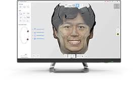
New Feature on iScan: Face Scan
You can now scan your face with the i500 via Medit iScan! In addition, you can also import 3D face data and bone data and align the various data sets for a more comprehensive final result.
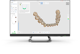
Capture Only the Data You Want to Keep: Smart Scan Filtering
We know the challenge and frustrations of capturing unnecessary soft tissue data while scanning. Ease your worries! With the smart scan filtering feature, you get to decide how much soft tissue data you’d like to capture while scanning, allowing you to choose the best option for each case.
Customize Your Scanning Workflow: Switching the Order of the Scan Stages
Do you prefer to scan the maxilla first before the mandible? Or the other way around? Whichever your preference, you now have the flexibility to choose the order of the scan stages to suit your working style and needs!
Medit Link V2.2 Release Notes
New Features -Web
The Medit Link homepage (meditlink.com) has been refreshed.
A new service landing page with information about the contents of the Medit Link service has been launched.
Membership service will be launched in the second half of 2020.
‘My Membership’ menu has been added for users to view their membership information.
The cloud storage capacity and other advanced features are offered based on the user’s membership plan.
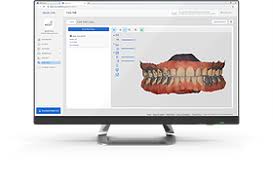
A new patient consultation feature ‘Case Talk’ has been added.
This patient consultation tool is available only for Clinic accounts and can be managed under the ‘Case Talk’ menu.
A list of all the cases created in the app, as well as scan data, can be found in the menu.
Communication with patients is possible by sharing a link to the Web Viewer via email or SMS.
My Information > Group Information: Users can check the record of members who have joined and left the group.
New Features – App
A button for Face Scan has been added to the form information window for the face scanning feature in iScan.
Face Scan files can be managed and used for orders in Medit Link.
The images captured in iScan can be edited using the Medit Link paint tool and can also be used when making orders.
The App Box has been added.
We have cooperated with Software Development Partners and Solution Providers to be integrated with Medit Link. You can install the available extension programs via App Box to complete the integration.
Experience a greater variety of workflows with the installed apps.
We will continue to add more apps in the App Box.
A banner space has been added at the top of the App Box page for partner introductions.
If you select exocad in the “Set Export Option” window, Medit Link will automatically create a compatible results file.
The raw scan files that were not uploaded can be reviewed and uploaded individually or in bulk.
The save path can be set to the local network drive.
However, the network drive will not be available in offline mode.
All users in the group can now be notified when a case has been completely processed.
Improvements
The UI of the Case Detail page has been improved.
Case history can be reviewed.
Case files can be viewed in the form of a list.
The transfer status of the file is shown by a progress bar with percentage display.
The 3D preview image has been improved by making the toolbar smaller so that the user can better focus on the image.
The support request window has been improved. Users can now choose the request type and select files to attach with the support request.
Available extension types to preview the attached files in have been added.
2D Image: png, jpg, jpeg, bmp, gif, tif, tiff
Video: mkv, mp4, avi, wmv
A button has been added to allow users to check the notices as well as to update details in ‘My Information’ window.
Previously, when a lab did not enter the shipping information, the cases in the order box could not be marked as complete because there was no delivery. The user can now change the status of these cases manually. This feature is not available in the In Box.
The names of the users who have deleted cases are now shown in the Trash Box.
Users can select the folder (save path) and input file name when exporting from Medit Link.
Bug Fixes
The following issues have been fixed:
Incorrect alignment of design file and scan results in the preview window.
Intermittent failure when synchronizing raw scan file.
Intermittent failure when importing files using the Case Converting Tool.
Partnership status being terminated without any restrictions.
Medit iScan V1.4 Release Notes
Improvement of UI/UX
The user interface is designed to be simpler and is categorized for the convenience of users.
Icon size has been reduced to make more space for the scan data.
If the scan stage meets the requirements, a check icon will be displayed to let the user know which stages are complete.
Tools that can be used only for the occlusion have been moved to the ‘Occlusion Scan Stage’.
‘Smart Data Cleaning’ which is related to data editing has been moved to the ‘Trim’ section.
‘Tools’ have been repositioned according to their functions.
The ‘Model Display Mode’ has been moved to the right-side bar – the touch screen controls are set to resemble a smartphone touch screen controls, and the ‘Pan’, ‘Zoom In/Out’, ‘Zoom Fit’ icons have been removed. They can be restored under settings.
The application can be used with a touch screen on the PC. For users running it on a laptop, trackpad keyboard controls have also been added.
Improvements in reliability map and post-processing algorithm have been made. The window which appears after the user clicks ‘Done’ previously showed three options for the 3D model. This has been simplified to two options for the convenience of users. You can also check the important conditions here before post-processing.
Smart Scan Filtering
This feature removes unnecessary soft tissue data which is the biggest challenge while scanning. Three filter options are available.
No Filtering
The previous basic filters (1,2,3) have been optimized and this filter is similar to the level one filter from the previous version. This is the basic filtering level and does not filter unwanted tissue. This option is useful for edentulous arch or plaster model cases.
Teeth + Gingiva
This filter removes soft tissue that interferes with the scan, leaving only the necessary gingiva. You can use this option for most general scan cases.
Teeth
This filter scans only the teeth. This option is effective for when you want to scan only the teeth as an additional scan, after scanning using the ‘Teeth + Gingiva’ filter.
Face Scan
You can scan your face with the i500, and also import 3D face data (STL, OBJ, PLY) and bone data (STL, OBJ, PLY) from an external source which have been converted from DICOM files taken via CT. The compatible data pairs can be aligned.
Measure
The ‘Measure’ tool allows you to measure the distance, angle, length, area, etc. of the 3D scanned data or section lines in iScan to check the progress of teeth preparation, height of the occlusion, as well as length of the teeth. The deviation result of two data sets can also be checked via the color map.
Reorder Scan Stages
Users can change the order of the scan stages.
Reposition Scan Data on The Occlusion Plane of Articulator
The scanned data is placed on the occlusion plane so that you can manually reposition it in case of incorrect positioning. In addition to that, the aligned result can be made compatible with the virtual articulator in exocad.
Improvement of A.I. Abutment Matching
You can save the margin line for abutments in the A.I. Abutment Matching dialog box. After you register the margin line, you can skip the process of drawing the margin line for each case in the dental clinic or lab. This saves a lot of time and unnecessary processing.
After the registered abutment is imported, the height is adjusted automatically or manually according to the scanned abutment. In this way, the saved abutments can be used for various cases.
Change Occlusion Target
Previously, occlusion alignment was only possible by using the maxilla and the mandible scan data in the ‘Scan Occlusion’ stage. Now, you can also use the pre-operation maxilla and the pre-operation mandible scan data.
Overview Stage
The ‘Overview’ feature has been added where you can view all the data simultaneously when you are not in any scan stage. Click the icon on the tree view on the left to hide or show each data set and use the slider to adjust the transparency.
Improvement of Replay
The ‘Scan Replay’ has been improved with options to choose scan steps. This helps the user view the scan replay video step-by-step.
Improvement of Post Processing Performance
Alignment of scan data is much more accurate in comparison to previous versions, resulting in better results with a smaller file size. Post-processing time has been reduced by about 20%.
Screen Capture
A screen capture feature and its corresponding tools have been added to the top right of the application window.
Improvement in Reliability Map
More conditions have been added to the Reliability Map for higher accuracy as compared to previous versions.
For users having difficulty in differentiating red and green colors, you can replace the green color with blue for the reliability map. Go to settings and ‘Reliability Map Color Mode’ to set the color.
Suitable Temperature for Calibration
The data accuracy increases if the temperature of the i500 during calibration is similar to the temperature while scanning.
The added feature to heat the device can be used in cases where the temperature of the i500 right before calibration is low.
Color Adjustment of Color Scan Data
The color processing algorithm and model rendering has been improved to display the scan data in a more natural way with more texture detail.
Changes and Improvement in ‘Settings’
The previously separate options have now been combined into single options of ‘Show Scanning Information’ and ‘Start Automatic Scan’.
The option to ‘Always scan in HD mode’ has been added for users who always scan in HD.
An option for the notification of external light detection (Beta) has been added. A strong light shining on the dentist chair can immensely affect the scan data, especially the anterior teeth data.
You can choose to display either three or six icons on the right side for ‘Model Control’.
The features ‘Use GPU’ and ‘Remove Soft Tissues’ have been removed as beta features and added as regular features. While using the GPU, a feature to check the basic specifications has been added and a message is displayed for each condition.
‘File Size’ and ‘Surface Roughness’ from the ‘Complete’ dialog box have been moved to the settings menu.
Improvement in Guide Messages
Guide messages for the user have been added and improved to make our product more user-friendly.
The user cannot get the best performance if the laptop power cable is not connected. A guide message is displayed in this case.
The scan strategy images and corresponding explanations have been improved so that the user can scan easily and correctly.
An ‘Overview’ has been added for the 3D model to help users operate the model conveniently.
Overall Improvements
Improved manual alignment in every step and a unified UI/UX.
Improved color bar algorithm in each stage to add the maximum and minimum value of deviation in extra boxes for calculation and to modify the maximum and minimum values on the color bar. Additionally, resolution control has been added to all the color bars to unify the UI/UX.
Improved algorithm for ‘Global Soft Tissue Filtering’ to prevent the tooth area from being deleted. It is no longer a beta version and has been changed to a regular feature.
Improved algorithm for ‘Scan Depth’, and the suitable filter values for each scan depth, are now automatically applied. It will automatically adjust to capture most of the data even when using deep scan depths, helping to scan deep areas without filtering out the necessary data.
Improved algorithm for ‘Scan Speed’ to calculate and display the scan speed more accurately than previous versions.
Improved ‘HD Camera’ algorithm to avoid shaky pictures taken with the i500 device. When you press the button, the camera automatically detects when there is minimum movement and maximum stability.
Improved saving algorithm for safe and secure storage of data.